Abstract: Background: Neuropathic osteoarthropathy (NO) commonly referred to as Charcot Foot, in patients with Diabetes mellitus, is a condition in which the bones, joints, and soft tissues of the foot and ankle are affected and involves inflammation in the initial phase. It further leads to reduced quality of life with high mortality rate and quality of life reduction. Several components interact to cause high incidence of fracture non-union, joint dislocation, foot deformity, and skin ulceration, increasing the relative risk of amputation. Annual incidence rates of 8.5/1000 per year have been reported. This rate has been increasing over the years with the availability of imaging modalities and reduced time in patient treatment
Objectives: To compare the clinical and radiological characteristics in diabetic patients with Neuropathic ulcers and to evaluate the severity of joint involvement in the Diabetic Neuropathy affected feet
Methods: In the year 2017 to 2018, a minimum sample size of 47 cases with diabetic neuropathy having osteoarthropathic changes satisfying inclusion and exclusion criteria and who have been admitted inpatient or treated on outpatient basis at our institution were selected. A cross sectional study on the samples was carried out. The aim of this study is to compare the clinical characteristics and radiographic changes in diabetic patients with neuroarthropathy and their outcome analysis.
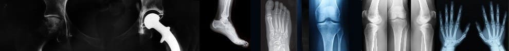
Results: In the total pool of 47 patients taken together, male predominance was found with maximum patients in the age group of 45-55 years. Diabetic patients with fore - foot ulcers had greater involvement than in mid – foot and Hind – foot ulceration when measuring the above mentioned angles with underlying neurological and vascular sign. Radiographic abnormalities were noted with changes mainly on Saggital and AP plains of the foot with predominance increased medial column height with reduction in calcaneal pitch and reduced Lateral calcaneal- 5th metatarsal angles in majority of the patients.
Conclusion: Findings in the sagittal plane differed than in Antero - posterior significantly when comparing patients on basis of area of involvement. The ultimate aim of the treatment is to prevent ulceration of the foot, and increase awareness among the patients having progressive deformity mainly in the sagittal plane. This will help to address the xray abnormalities and plan for further reconstructive CN surgery re-establishing the normal anatomy.